BackTable / ENT / Article
Thyroid Imaging: What is the “Gold Standard” for Thyroid Nodules?
Taylor Spurgeon-Hess • Updated Aug 29, 2024 • 337 hits
With continuous technological advancements, numerous thyroid imaging modalities exist to capture a clear picture when it comes to looking at thyroid nodules. What role do each of these modalities play in prioritizing both efficiency and comprehensiveness?
This article features excerpts from the BackTable ENT Podcast. We’ve provided the highlight reel here, and you can listen to the full podcast below.
The BackTable ENT Brief
• Thyroid ultrasound imaging effectively captures the majority of information needed for working with thyroid nodules, and remains widely available to patients without compromising on efficacy.
• Resolution of discrepancies between imaging and biopsy relies on individual conversations and the patient’s personal risk aversion. With patient compliance and moderate risk tolerance, the course of action often includes a six-month reassessment and follow-up.
• CT scans of thyroid nodules can provide additional information in cases where the ultrasound or FNA biopsy results do not provide a clear enough picture.
• Other thyroid imaging modalities may not be first-line, but can assist in diagnosing thyroid nodules not easily noticed or palpated.

Table of Contents
(1) Thyroid Ultrasound Utility
(2) Handling Thyroid Imaging Discrepancies
(3) The Role of PET and CT Scans of Thyroid Nodules
Thyroid Ultrasound Utility
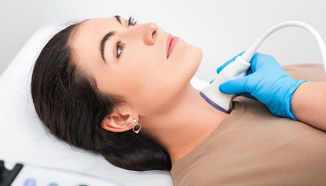
Thyroid ultrasound imaging is both inexpensive and effective in obtaining an image of a thyroid nodule. While not a new technology, advancements allow today’s ultrasound to detect almost any sign of malignancy. This thyroid imaging modality shines when it comes to identifying abnormal lymph nodes. Dr. Goldenburg recommends that, no matter the size of the nodule, both sides of the neck should be scanned in the ultrasound.
[David Goldenberg MD]
Well, the imaging modality of choice for anything thyroid, whether it's in the thyroid or in the neck is the ultrasound, the good old-fashioned, inexpensive, non-radiating ultrasound. And it's an interesting point. If they come in with their MRI, that's wonderful. I don't really like it when they were sent specifically for an MRI for a five-millimeter nodule, but it happens sometimes again, you know, when patients are referred into our practice specifically for this, oftentimes they come in with all kinds of imaging, but ultrasound is the tried, true, tested imaging modality.
[Ashley Agan MD]
Do you do any ultrasound, in your clinic or are most of your ultrasounds done in your radiology department?
[David Goldenberg MD]
I am fully credentialed to do ultrasounds and I'll do them every now and again myself, if I think it will add something. A lot of times the patients are sent to radiology, just because of timing. Just takes a while and sometimes patients will come in with an ultrasound already performed.
So there's really, it's a whole bag of, but certainly in our offices, we'll do an ultrasound if we think that it will add something, yeah. Labs typically TSH, if it hasn't been done, is the only thing you really need. And that's just to rule out a, you know, a toxic nodular goiter, incredibly rare in this day and age.
[Gopi Shah MD]
I assume that usually if there's a concern for a thyroid nodule, and you send them for an ultrasound thyroid you also have them scan the neck, both sides, one side?
[David Goldenberg MD]
Gopi. That's a really interesting point. I teach my residents that a thyroid ultrasound must include both sides of the neck. Because regardless of the nodule size, regardless of how benign it looks, if the patient has abnormal-looking lymphadenopathy that trumps everything. Ultrasound is excellent for looking for abnormal lymph nodes in the lateral neck.
It is not sensitive for looking for lymph nodes in the central compartment while the thyroid gland is in situ. So, you know, if they see something, that's also suspicious, but if they don't see something, it doesn't mean that it's not there. Ultrasounds are so good nowadays that we can get an inkling of the suspicion for malignancy in a thyroid nodule based on findings on ultrasound. So if someone comes in with a cystic lesion, purely cystic, the chances of this being malignant are very, very low. On the other hand, if a patient comes in with a nodule, which has, oh, microcalcifications, a hypoechoic irregular margins, taller rather than wide, mixed cystic solid component. All of these things say to me that the chances that this nodule regardless of size may be malignant. And, we'll probably take that to the next diagnostic level.
Listen to the Full Podcast
Stay Up To Date
Follow:
Subscribe:
Sign Up:
Handling Thyroid Imaging Discrepancies
When discrepancies between imaging and fine needle aspiration biopsy occur, a multitude of factors dictate the doctor-patient conversation that follows. These factors include patient compliance, preference, and risk aversion. In years past, physicians ordered lobectomies to prevent missing something malignant, but these procedures are irreversible and sometimes unnecessary. In order to avoid the overuse of lobectomies, Dr. Goldenburg recommends scheduling an ultrasound six months out to reassess when working with patients likely to return to the office for a follow-up.
[David Goldenberg MD]
…So what do you do about discrepancies? What do you do when things don't line up? So in this day and age, this is a conversation that you have with a patient. And although they taught us in medical school to speak with patients, I don't think maybe they're teaching it now, but not all patients are created equal. Some of them have an understanding. Some of them don't, some of them don't want to have an understanding. “You're the doctor, you decide.” To which I usually answer, “It's your neck, you decide.” But it's a conversation and we do it together.
So we have to ascertain the patient's risk aversion. We have to ascertain whether they can sleep at night, whether they're an upstanding citizen, who's going to come back for another ultrasound or a biopsy or disappear in the wind only to come back five years later with an anaplastic thyroid cancer. These are things I've all seen. So all of these things, it's, it's not just the FNA.
It's not just that conversation has to be had. I think you'll never go wrong if you bring the patient back for an ultrasound in six months. Never. I also don't think that you're wrong if you do a lobectomy on a patient like that, because they can't sleep at night, thinking that there's a chance that maybe it's a cancer.
Certainly years ago, we did a lot more lobectomies. We know how to do that. We know how to do it well. The patients will be great. It's not the wrong answer either. Last week I had a patient who came in with a biopsy in an outside institution that was, sent out for Afirma, 50% chance of malignancy suspicious.
I always bring the path in to our institution. I have a patient who came in with a fine needle aspiration biopsy, an outside institution that was Afirma suspicious, 50% chance of malignancy sent to me for a lobectomy.
I always ask that pathology be brought to our institution because I always think a second opinion by people who experienced it. It's not never a bad idea. Well, our people said there's insufficient cells to call it anything. So I sent her for another biopsy. My new biopsy came back completely benign. So what do we do?
Do we believe the old one and give her a lobectomy, which is probably unnecessary. Or do we wait? So it's a discussion I had with a patient. I try not to push patients one way or another, but if they now, you know, I'll say, “Listen, if I think you're doing the wrong thing, I'll say to you, I think you're doing the wrong thing.”
In this case, we're going to get her ultrasound in six months. Even if this is a papillary, these are slow-growing cancers. We all know that. The main thing is not letting the patient get lost and having a follow-up system so that you keep an eye on it.
The Role of PET and CT Scans of Thyroid Nodules
Many patients presenting to an ENT go in for PET or CT thyroid nodule maging ordered for unrelated conditions and end up discovering the thyroid nodule serendipitously. In these cases, the imaging provides a picture of the nodule, but ultrasound and/or fine needle aspiration biopsy may still be needed to supplement. Although not routine, if a physician questions the need for surgery, CT thyroid scans can assist in determining the proper course of action.
[Gopi Shah MD]
And then in terms of like workup for your nodules, you know, for some of the cases that aren't as clear cut, the example that Ash gave was a great one. Do you ever use a PET scan or is there a role for these other imaging modalities and how do you use them?
[David Goldenberg MD]
That's a wonderful question because we see this. So the vast majority of people with PET scans are people who come in with a PET scan for another malignancy and they find a solitary FDG avid nodule, and those are actually considered suspicious and need to be worked up and they are.
So the patients come in with a PET CT scan for another reason, if there's a solitary FDG avid nodule that needs to be worked up, usually they get a fine needle aspiration biopsy on the ultrasonic guidance.
In those cases, they don't need a neck CT because the neck is covered by the PET. And overwhelming, if the thyroid cancer is FDG avid, and it's a cancer, it's usually more aggressive because it means it's probably not iodine avid and the opposite. So I do remember a couple of patients who accidentally came in the young, you know, sometimes a patient will not be sent to us or to even endocrinology by a PCP because they hear the word cancer and they send it to oncology and they do a PET scan probably unnecessarily. And you see it. So this is kind of anecdotal if you will. So no, we don't send for, for our thyroid nodule for a PET scan. if I'm doing surgery on a patient that already has a thyroid cancer. Or if, a thyroid nodule, like if someone comes in with a thyroid nodule and they are complaining of compressive symptoms, my rule is that I have to be convinced that it is the thyroid nodule that is causing their compressive symptoms.
The reason being, there's an old saying, you break it, you buy it. So once you've operated on the patient, they're yours for life. And if you're taking out half their thyroid gland for no good reason, you're not going to make them happier because thyroidectomy doesn't typically cure reflux. So I'll get a CAT scan on someone who is complaining of compressive symptoms.
And I will go over the scan together with the patient, say to them, “You know what, you're right. Your massive thyroid nodule is compressing your esophagus or pushing on your trachea. Let's take out below.” or I'll say to them, “You know, your thyroid is really not that large. We have to look for another reason to explain your compressive symptoms.”
If it's a thyroid cancer, I reserve the right to do a CAT scan with contrast if I think it will help my surgical planning. Usually if there's extensive neck disease, I say I reserve the right, because in certain places, nuclear medicine, gets up in arms over the use of contrast. I think it's unwarranted personally.
We do what's right for the patient. If I think a CAT scan will help, but it's not a routine thing that I'll do for any old thyroid nodule.
[Ashley Agan MD]
And the concern is timing for post-op radioactive iodine. Is that right? They're worried that that's going to delay that treatment if they need it.
[David Goldenberg MD]
Yeah. That's the concern. Yeah, I don't buy it. First of all, it takes awhile to get the nuclear scan or the iodine treatment, at least in our neck of the woods. I'm sure the iodine from the CAT scan has washed out long ago. But in any case, my ability to perform surgery is at that point, the more important thing. It's a surgically treated disease, right?
Podcast Contributors
Dr. David Goldenberg
Dr. David Goldenberg is a professor and the chair of the department of otolaryngology - head and neck surgery at Penn State in Hershey, Pennsylvania.
Dr. Ashley Agan
Dr. Ashley Agan is an otolaryngologist in Dallas, TX.
Dr. Gopi Shah
Dr. Gopi Shah is a pediatric otolaryngologist and the co-host of BackTable ENT.
Cite This Podcast
BackTable, LLC (Producer). (2021, November 2). Ep. 35 – Thyroid Nodules [Audio podcast]. Retrieved from https://www.backtable.com
Disclaimer: The Materials available on BackTable.com are for informational and educational purposes only and are not a substitute for the professional judgment of a healthcare professional in diagnosing and treating patients. The opinions expressed by participants of the BackTable Podcast belong solely to the participants, and do not necessarily reflect the views of BackTable.


